| dc.contributor.author | Demircan, Mehmet | |
| dc.contributor.author | Gürünlüoğlu, Kubilay | |
| dc.contributor.author | Gözükara Bağ, Harika Gözde | |
| dc.contributor.author | Koçbıyık, Alper | |
| dc.contributor.author | Gül, Mehmet | |
| dc.contributor.author | Üremiş, Nuray | |
| dc.contributor.author | Gül, Semir | |
| dc.contributor.author | Türköz, Yusuf | |
| dc.contributor.author | Taşcı, Aytaç | |
| dc.contributor.author | Gürünlüoğlu, Semra | |
| dc.date.accessioned | 2022-03-10T13:27:28Z | |
| dc.date.available | 2022-03-10T13:27:28Z | |
| dc.date.issued | 11.12.2020 | en_US |
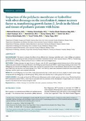
| dc.identifier.citation | Demircan, M., Gürünlüoğlu, K., Gözükara Bağ, H. G., Koçbıyık, A., Gül, M., Üremiş, N., ... & Taşçı, A. (2021). Impaction of the polylactic membrane or hydrofiber with silver dressings on the interleukin-6, tumor necrosis factor-α, transforming growth factor-b3 levels in the blood and tissues of pediatric patients with burns. Turkish Journal of Trauma and Emergency Surgery, 27(1), 122-131. | en_US |
| dc.identifier.uri | DOI: 10.14744/tjtes.2020.30483 | |
| dc.identifier.uri | https://hdl.handle.net/20.500.12899/595 | |
| dc.description.abstract | BACKGROUND: We aimed to evaluate the effects of two different burn dressings, hydrofiber with a silver (HFAg) and polylactic membrane (PLM), on altering the levels of important biomarkers Interleukin-6 (IL-6), Tumor necrosis factor-α (TNF-α), Transforming growth factor-β3
(TGF-β3 ) in blood and burnt tissue in children with second-degree burns.
METHODS: Children between the ages of one to 16 years, with 25–50% second-degree partial-thickness burns of the total body surface area were included in this study. Patients in the PLM group were dressed with PLM in a typical way according to the manual. The HFAg group was dressed with HFAg and a sterile cover. During and at the end of the 21-day treatment, blood and skin tissue samples were taken from the two burn and control groups. IL-6, TNF-α, and TGF-β β3 levels were evaluated in blood and tissue samples from all groups, and the results were analyzed statistically.
RESULTS: In the PLM group, IL-6 and TNF-α levels decreased early days in both serum and tissue samples to reach normal ranges compared with the HFAg group. In the PLM group, TGF-β3
levels were elevated than in other groups for two weeks.
CONCLUSION: In this study, we found that PLM controls inflammation earlier in both systemic and burn tissue. We also found that PLM increased the level of TGF-β3 , which may be associated with the prevention of the development of hypertrophic scar in the burn wound, in the blood and burn tissue during this study | en_US |
| dc.language.iso | en | en_US |
| dc.relation.ispartof | Turkish Journal of Trauma and Emergency Surgery | en_US |
| dc.rights | info:eu-repo/semantics/openAccess | en_US |
| dc.subject | Burns | en_US |
| dc.subject | children | en_US |
| dc.subject | dressing | en_US |
| dc.subject | IL-6 | en_US |
| dc.subject | hydrofiber with silver | en_US |
| dc.subject | polylactic membrane | en_US |
| dc.subject | TNF-α TGF-b | en_US |
| dc.title | Impaction of the polylactic membrane or hydrofiber with silver dressings on the interleukin-6, tumor necrosis factor-α, transforming growth factor-b3 levels in the blood and tissues of pediatric patients with burns | en_US |
| dc.type | Article | en_US |
| dc.authorid | 0000-0002-4668-9603 | en_US |
| dc.department | MTÖ Üniversitesi, Tıp Fakültesi, Temel Tıp Bilimleri Bölümü | en_US |
| dc.institutionauthor | Gürünlüoğlu, Semra | |
| dc.identifier.doi | 10.14744/tjtes.2020.30483 | |
| dc.identifier.volume | 27 | en_US |
| dc.identifier.issue | 1 | en_US |
| dc.identifier.startpage | 122 | en_US |
| dc.identifier.endpage | 131 | en_US |
| dc.relation.publicationcategory | Makale - Uluslararası Hakemli Dergi - Kurum Öğretim Elemanı | en_US |
| dc.identifier.pmid | 33394485 | |
| dc.identifier.scopus | 2-s2.0-85099428890 | en_US |
| dc.identifier.wos | WOS:000604992400021 | en_US |
| dc.identifier.wosquality | Q4 | en_US |
| dc.indekslendigikaynak | Web of Science | en_US |
| dc.indekslendigikaynak | PubMed | en_US |